Many common resistance-training exercises designed to isolate specific muscles don’t account for the separate skeletal structures and how they are designed to move as a single unit. In this fourth installment of our functional anatomy series, we examine the integrated actions of the four joints that make up the shoulder complex. Specifically, we’ll focus on how the separate joints are designed to function as an integrated unit and the common exercises that could be causing injuries. We’ll also highlight strategies for improving the stability, mobility and strength of this oft-injured body part.
The Structures of the Shoulder
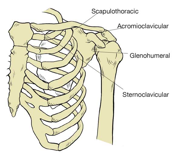
The skeletal system of the body is organized into two different components: the axial skeleton, which is composed of the spine, skull and rib cage, and the appendicular skeleton, which includes the pelvis, legs, shoulders and arms. The shoulder is comprised of four distinct joints and is the foundation of movement for the arms (Neumann, 2010):
- The sternoclavicular (SC) joint, where the proximal end of the clavicle floats on the sternum and first rib, is a relatively small joint that connects the shoulder and arm directly to the rib cage of the axial skeleton.
- The acromioclavicular (AC) joint is where the distal end of the clavicle aligns with the acromion process of the scapula.
- The glenohumeral (GH) joint is where the spherical head of the humerus lies in the concave surface created by the glenoid fossa of the scapula; it is the joint most people think of when considering the shoulder.
- The scapulothoracic (ST) joint isn’t technically a joint (which is defined as the point where two bones connect), but is the location where the scapula floats on the posterior surface of the rib cage. Each individual scapula is connected via 17 muscles responsible for allowing mobility to support the movement of the humerus, as well as creating the stability necessary for optimal posture and to support overhead movements.
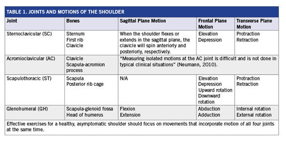
The ball-and-socket structure of the GH joint allows a significant range of motion in all three planes. The ST and SC joints allow motion primarily in the frontal and transverse planes, while the AC joint allows only a limited amount of motion, which is critical for the optimal function of the other joints (Table 1). Individually, each of these joints allows a specific range of motion, but when functioning as an entire unit the shoulder becomes one of the most mobile parts of the human body. The paradox of the shoulder complex is that it needs to have the stability required to prevent unwanted motion, while also having the mobility necessary for many dynamic movements (Wilk, Meister and Andrews, 2002). If any of the four joints is out of alignment it will affect the ability of the other joints to perform their requisite functions to allow optimal mobility or provide necessary stability.
Understanding Movement of the Shoulder Joint
The shoulder can move in all three planes of motion—sagittal, frontal and transverse—and many common exercises focus on movement of the shoulder in one of these planes at a time. However, when selecting exercises to enhance shoulder strength, it is important to consider that there is a fourth plane of motion created by the way the glenoid fossa of the scapula sits relative to the thoracic rib cage. In a healthy shoulder, the concave socket of the glenoid fossa points in an angle that is approximately 35 degrees anterior to the frontal plane (Neumann, 2010). This fourth plane of motion is often overlooked in many shoulder exercises, which could potentially be a cause of many injuries. The position of the glenoid fossa means that overhead movements should be neither wholly in the sagittal plane nor in the frontal plane, but should be somewhere in the middle, where the elbows are pointing approximately 30 to 45 degrees from the center front of the body (at about the 10 o’clock and 2 o’clock positions).
Overhead Presses and Front Raises
Shoulder presses done in the frontal plane, with the elbows pointed at 9 and 3 o’clock, could cause an injury to specific shoulder muscles. When the humerus is held in an externally rotated position to perform an overhead press in the frontal plane, the deltoid tuberosity can impinge the long head of the biceps brachii and the supraspinatus muscle between the coracoid and acromion processes, respectively. In addition, as the supraspinatus is compressed between the humerus and acromion process, it changes the length-tension relationships of the other rotator cuff muscles, which can inhibit their ability to effectively stabilize the humerus when the arm is extended overhead. Try this: Hold your arms out in the frontal plane and perform a few overhead presses. You should feel a slight impingement about 75 to 80 percent of the way through the motion. Now move your arms to the mid-way point between the sagittal and frontal planes and try a few presses. You should feel how your arm can extend all of the way overhead without any restriction. With the arm overhead in the scapular plane, the serratus anterior, lower trapezius and upper trapezius are in an optimal position to stabilize the ST joint, which is a much safer way to perform overhead presses.
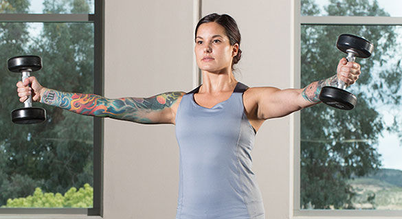
Performing front raises while holding the hands in a palms-down, pronated position creates internal rotation at the GH joint, which can reduce space in the anterior compartment of the joint, impinging the long head of the biceps brachii. A more effective way to strengthen the anterior deltoids is to hold the hands in a neutral position with the palms facing the midline of the body or to perform V-raises (see the workout for shoulders below).
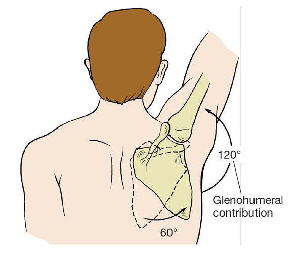
The term “elevation” is used to describe movement of the arm overhead without identifying the specific plane of motion. Movement of the arm into the overhead position is produced by muscles that can be organized into three groups: (1) muscles that abduct and/or flex the humerus at the GH joint, (2) muscles that control upward rotation of the scapula at the ST joint and (3) the rotator cuff muscles that control both mobility and stability at the GH joint (Neumann, 2010). As the arm is elevated above the height of the shoulder, the glenoid fossa should move under the head of the humerus to support the weight of the arm (and whatever it might be holding) as it extends overhead; the technical term for this is scapulohumeral rhythm. In a healthy shoulder, as the deltoids contract to move the humerus overhead in the scapular plane, the muscles of the rotator cuff, specifically the supraspinatus, infraspinatus, teres minor and subscapularis, pull the head of the humerus into the glenoid fossa to create stability as the arm moves into the fully extended, overhead position. In an injury-free shoulder, there should be a 2:1 ratio of movement of the humerus to the scapula. As the humerus moves to a position where it is 120 degrees relative to the body, the scapula will upwardly rotate (the point of reference is the glenoid fossa) up to 60 degrees to support the arm (American Council on Exercise, 2015).
The Role of the Shoulder Muscles
During normal movements, muscles contract as a reflex to provide the force required to either stabilize a joint to prevent unnecessary motion or to cause a specific segment of the body to move. Whether the function is to provide stability or initiate motion, an essential component of a muscle’s ability to function properly is its resting length. If a muscle is held in either a shortened or lengthened position for an extended period of time, its ability to function properly will be altered. When the resting length of a muscle changes, both the position of the bones to which it’s attached as well as the structure of the joints where it influences motion will be changed as well (American Council on Exercise, 2015).
Many people have poor upper-body posture, with shoulders that are slumped and rounded forward. This can drastically change the length-tension relationships of the muscles; specifically, the second group of muscles that control upward rotation of the scapula (trapezius, levator scapula, rhomboid and serratus anterior) and are responsible for moving and stabilizing the scapula to maintain optimal glenohumeral rhythm. So, does a change in muscle length change the alignment of a joint or does a misalignment of a joint change the length of involved muscles? “It’s not exactly clear,” says Gray Cook, a physical therapist and creator of the Functional Movement Screen, “but we do know that poor posture or alignment changes the information relayed to the central nervous system, which can inhibit muscle activity.
“What is important,” Cook continues, “is to first strengthen the muscles responsible for stabilizing the shoulders before focusing on the muscles that create movement at the joints.”
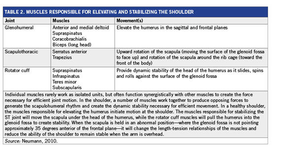
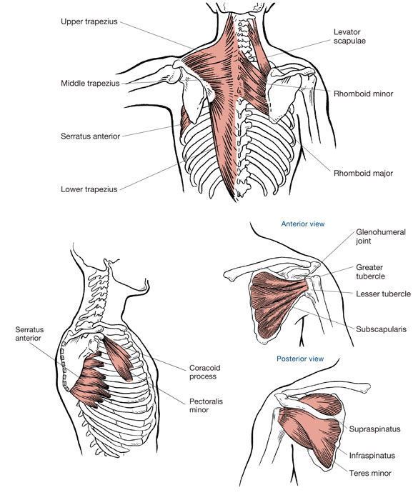 All of the muscles responsible for stabilizing and moving the shoulder should be trained in a way that improves their ability to function, allowing the scapula and humerus to move together as a single integrated unit. The anterior, medial and posterior heads of the deltoid help abduct the humerus away from the midline of the body and are some of the most visible muscles of the shoulder complex. As a result, the traditional overhead press focuses the movement in the frontal plane in an effort to maximize the recruitment of these muscles. However, successful, pain-free overhead movements with the shoulder rely on the deeper muscles of the rhomboid, serratus anterior and levator scapula, as well as the superficial trapezius, to work together to move and stabilize the scapula to create a stable foundation as the arm is raised overhead. The deltoids will generate the force to move the humerus; however, the scapular stabilizers are responsible for moving the cup of the glenoid fossa as the rotator cuff muscles hold the head of the humerus stable in the joint (with the joint surface of the glenoid fossa pointed 30 degrees anterior of the frontal plane). As the arm moves overhead and the scapula rotates upward to support the weight of the limb, the muscles of the rotator cuff, the supraspinatus, infraspinatus, teres minor and subscapularis work to pull the head of the humerus into the socket created by the glenoid fossa. Rounded, protracted shoulders can change the length-tension relationship of all of these muscles, which will significantly alter how the joint functions when the arm is raised, especially if the elbows are held in the frontal plane, as they are during traditional overhead presses. If the muscles of the shoulder cannot properly stabilize the scapula and control motion of the humerus to create optimal scapulohumeral rhythm as the arm moves above shoulder height, impingement could occur and be a potential mechanism for injury. Effective exercise program design for the shoulder should first create stability of the ST joint, restore the length-tension relationships of the muscles responsible for controlling the scapula and, finally, generate the force to both create stability of the GH joint and move the arm overhead (Table 2).
Exercise Strategies for Improving the Stability and Strength of the Shoulder
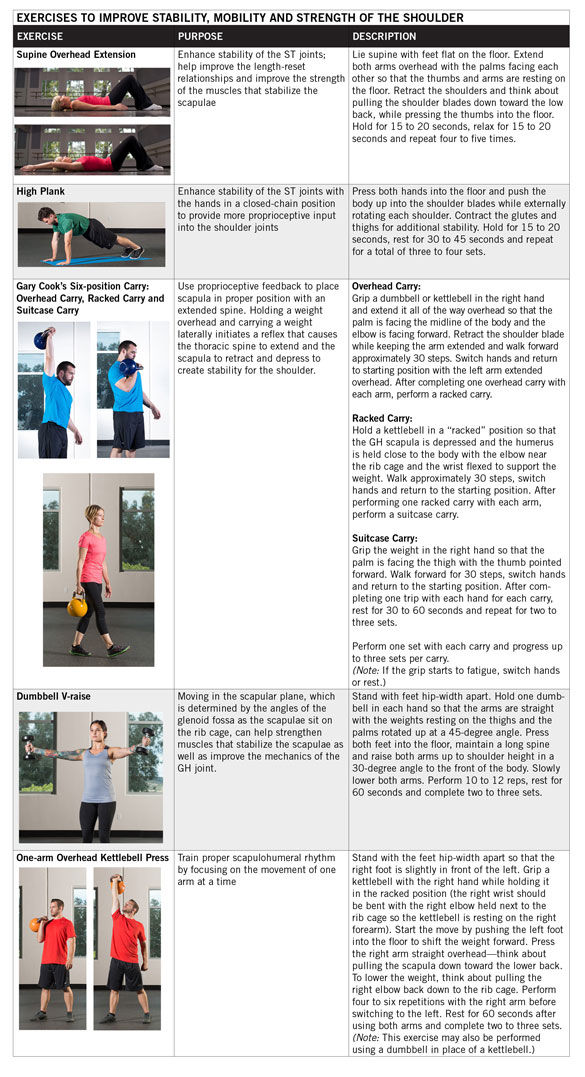
The first step in developing a strong, functional shoulder is improving the stability of the ST joint and restoring the length-tension relationships of the muscles responsible for moving and stabilizing the scapula. Isometric contractions that maintain consistent force can help improve the strength of the middle and lower trapezius as well as the rhomboids, which is important for creating ST joint stability. Lying supine with the arms raised overhead so that the palms are facing one another can help teach a client how to engage the scapular stabilizers with the humerus in a flexed, overhead position. And the high plank, which is a closed-chain (hands-on-floor) exercise, can help to develop shoulder stability and strengthen the scapular stabilizers and rotator cuff muscles.
Once the initial stabilization is improved, Cook suggests training the entire shoulder complex using exercises that enhance the ability of the muscles to create stability as an automatic reflex. “The muscles responsible for dynamic stability of the scapula and ST joint are reflex dependent,” explains Cook. “When placed in the correct position, the body will know how to create dynamic stability as an automatic response. If your alignment is good and your body position is where it needs to be, your brain will reflexively fire to give the joint necessary integrity.”
Cook’s go-to exercise series for improving the stability and strength of the shoulder is the Six-position Carry, which requires an individual to carry a kettlebell with three specific holds for each arm: the Overhead Carry, the Racked Carry and the Suitcase Carry (see workout below). Cook prefers the kettlebell because it requires a strong grip and the muscles responsible for grip strength have direct “feed-forward neurology” to the muscles responsible for moving and stabilizing the shoulder.
When it comes to helping clients improve their strength, Cook urges trainers to focus on allowing clients to develop the necessary reflexes by using integrated movement patterns rather than isolated motions. “We should coach position,” Cook argues. “If you have to tell somebody to engage their muscles, what is going to happen when they get in a real-world situation and you’re not there to help them execute the pattern?”
Your role as a health and fitness professional is to help your clients achieve their goals in the safest, most time-efficient manner possible. As such, understanding how the four joints of the shoulder are designed to work together can help you identify the safest, most effective strategies to help clients improve their upper-body strength. When it comes to designing a workout, your objective should be to help clients develop reflexive strength so they can be successful, even when you aren’t standing right there coaching an exercise. After all, empowering clients to be successful on their own is one of the most important results you can deliver.
References
American Council on Exercise (2015). ACE Medical Exercise Specialist Manual. San Diego, Calif.: American Council on Exercise.
Neumann, D. (2010). Kinesiology of the Musculoskeletal System: Foundations for Rehabilitation (2nd ed.). St. Louis, Mo.: Elsevier.
Wilk, K., Meister, K. and Andrews, J. (2002). Current concepts in the rehabilitation of the overhead throwing athlete. The American Journal of Sports Medicine, 30, 1, 136-151.


 by
by