As a fitness professional and an exam candidate, there is no way of getting around the fact that you need to know your anatomy! It is important to understand how the body moves and how muscles work together to generate movement. In other blogs, we looked at how to study anatomy, muscles that move the scapulae, the muscles that move the arm and the muscles of the core. Here, we will look at the muscles of the hip, knee and ankle joints.
Hip Joint
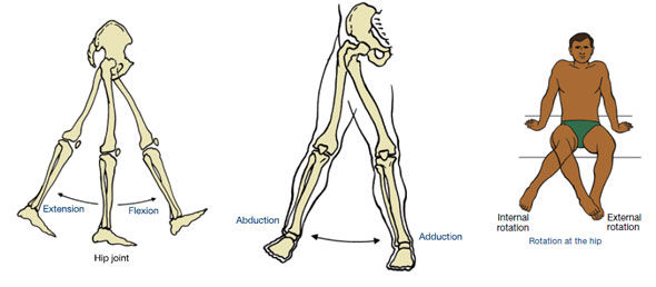
- The hip joint is created between the femur (thigh bone) and the acetabulum of the pelvis (socket of the hipbone).
- Similar to the shoulder joint, it is a ball and socket joint that has many actions.
|
Action of the Hip |
What the Action Looks Like (Move Your Body!) |
Primary Muscles |
|
|
Hip Flexion |
Lift your thigh upward in front of your body |
|
|
|
Hip Extension |
|
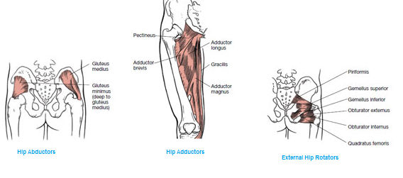
Hip Extensors – hamstrings (focus on biceps femoris) and gluteus maximus |
|
|
Hip Abduction |
Lift your leg out to the side, or from a squatting position, knees falls out to the side |
Hip Abductors - gluteus medius and minimus |
|
|
Hip Adduction |
From a position of hip abduction, lower your thigh to the anatomical position |
Hip Adductors (know them as a group called the hip adductors) |
|
|
Internal Rotation of the Hip |
Rotate your leg in toward the midline of your body |
Because of the anatomical configuration of the hip, there are no true primary internal rotators of the hip. Muscles that play a role in internal rotation when the hip is first flexed to 90 degrees are the tensor fasciae latae, adductors longus and brevis, pectineus and the anterior fibers of gluteus medius and minimus. |
|
|
External Rotation of the Hip |
Rotate your leg out away from the midline of your body |
External rotators (know them as a group called the external hip rotators); focus on piriformis because of its role in sciatica |
Knee and Ankle Joints
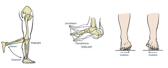
- The knee joint consists of the end of femur bone connecting with the top of the tibia and fibula.
The two main actions of the knee are flexion and extension.
- The ankle joint consists of the distal ends of the tibia and fibula and the talus.
- The main actions of the ankle are plantarflexion, dorsiflexion
- The subtalar joint (articulation between the talus and calcaneus) allows inversion and eversion of the foot
|
Action of the Knee and Ankle |
What the Action Looks Like (Move Your Body!) |
Primary Muscles |
|
Knee flexion |
Bend your knee |
Knee flexors – hamstrings, focus on biceps femoris |
|
Knee extension |
Straighten your knee |
Knee extensors – quadriceps, focus on rectus femoris |
|
Ankle plantarflexion |
Point your toes with the foot off of the ground, or when standing, lift your heels off the floor |
Plantarflexors: (know them as a group called the plantarflexors); focus on gastrocnemius and soleus |
|
Ankle dorsiflexion |
Lift your toes up off the floor toward your shin |
Dorsiflexors: (know them as a group called the dorsiflexors); focus on anterior tibialis |
|
Ankle inversion |
Pull the foot toward the midline (ankle rolled out) |
Anterior tibialis |
|
Ankle eversion |
Pull the foot away from the midline (ankle rolled in) |
Personeus longus and peroneus brevis |

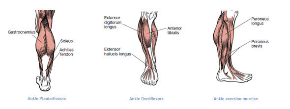
A helpful way to learn anatomy is to move and mimic the actions for the muscles you are learning that week. Look at the picture of the muscle, find it on your body, and picture how it is contracting as it produces its associated movement or movements. That is, contract the muscle you are reviewing and complete the different actions that the muscle is capable of making.


 by
by 




 by
by