The abdominal muscles, specifically the rectus abdominis, are often trained in an inefficient manner that may not actually improve their mechanical ability to function as a component of an integrated system responsible for producing and controlling human movement. While many clients want to do crunches in an attempt to sculpt a “six-pack,” the only time the rectus abdominis flexes the spine is when the body is lying on the ground (in other words, when a person is doing crunches). This, of course, begs the question: Are crunches really the most effective way to strengthen this muscle? Closely examining how the abdominal muscles are designed to function during upright movement patterns makes it clear that that a number of traditional exercises, such as crunches, should not be the foundation of an effective strength-training program for muscles that control movement at our body’s center of gravity.
The first in an exclusive new ACE series covering the major muscles of the body, this article describes how the abdominal muscles are designed to work as an integrated system and how to identify strategies for using exercise to achieve optimal function.
Understanding Movement
Creating an exercise program requires an understanding of how the body is designed to move. Exercise is not a series of separate, discrete muscle actions, but rather a function of movement created by many muscles working together simultaneously. Proper exercise selection for workout programs requires knowledge of how muscles function as an integrated system, so you can help clients train them to produce the right amount of force at the right time.
The human body is designed to move and the foundational pattern of human movement is the gait cycle; therefore, the musculoskeletal structures of the human body are designed to be most efficient when standing upright on the ground, not lying on the floor. The skeletal and muscle structures in our bodies create kinetic energy from gravity and ground-reaction forces to conserve metabolic energy. This means that, as a mechanical structure, the human body functions most efficiently when walking or running across the ground.
The traditional approach of understanding human anatomy for the purpose of designing exercise programs is based on the assumption that muscles function independently to perform separate, isolated actions. A more appropriate way to describe anatomy as it relates to human movement is to recognize that all muscles work together to accomplish a specific task. Additionally, understanding how anatomical structures function as components of a completely integrated system is necessary for designing exercise programs that can help clients achieve their aesthetic and performance-based goals.
Controlling Movement at the Center of Gravity
The word “core” is commonly used to describe the muscles that control motion at the pelvis, femurs, rib cage or lumbar spine. This is an incomplete understanding, however, because a number of muscles can influence motion at these structures that are not traditionally considered part of the body’s “core.” For example, the long head of the biceps attaches to the supraglenoid tubercle, and the short head attaches to the coracoid process of the scapula, which sits on the thoracic rib cage. If the biceps remain in a state of contraction, it could pull the scapula out of position, which then changes the position of the thoracic spine. In another example, consider the superior attachments of the gastrocnemius on both the medial and lateral condyles of the femur. If the gastrocnemius remains in a state of contraction, it can influence movement of the femur and joint mechanics at the hip.
Researcher Dr. Stuart McGill describes the core as being “composed of the lumbar spine, the muscles of the abdominal wall, the back extensors and quadratus lomborum. Also included are the multijoint muscles, namely, latissimus dorsi and psoas that pass through the core linking it to the pelvis, legs, shoulders and arms” (McGill, 2010). A separate model described by Bergmark (1989) identifies core muscles with attachments to the lumbar vertebra as “local,” while muscles with attachments to the hips and pelvis are “global.” The local muscles control intersegmental stability of the lumbar spine, while the global muscles influence orientation of the spine and help control the forces that cause it to move (Hibbs et al., 2008). In yet another model, Neumann (2010) describes the abdominal muscles as being organized into layers: superficial, intermediate and deep. Regardless of the specific model, abdominal muscles function as the transmission of the body, because they are responsible for transferring forces generated from the ground, through the legs and trunk and, ultimately, out through the upper extremities.
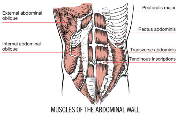
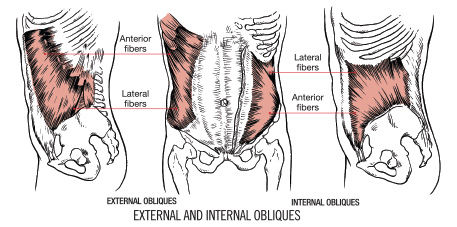
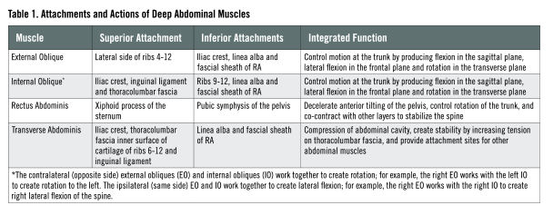
The external and internal obliques, rectus abdominis (RA) and transverse abdominis (TVA) are all considered part of the deep layer in Neumann’s model or as global muscles in Bergmark’s research, and comprise the abdominal wall as described by McGill. The muscles of the abdominal wall are layered against one another like the individual layers of a sheet of plywood. Because of the way the muscles are structured, they are not capable of working in isolation and will instead contract together as one unit during upright movement. These deep muscles work together to control motion of the spine, rib cage and pelvis. Table 1 describes the attachments and actions of the deep abdominal muscles.
Effective training for abdominal muscles requires using movements that integrate the hips, trunk and shoulders to efficiently distribute the forces created by gravity, ground reaction and momentum during upright movements. Dr. Gary Gray, a physical therapist and founder of the Gray Institute, teaches courses on anatomy, biomechanics and how to improve physiological function. "The abdominals can be controlled by the brain to flex the spine when you're lying on the ground,” explains Dr. Gray, “but this is not how they actually work. The multiple layers of the abdominals are lengthened in all three planes as the rib cage and pelvis rotate opposite one another during upright movement in gait. This lengthening motion turns the muscles on reflexively, which is their natural way of functioning.”
You Learned Everything You Needed to Know About Your Core Before You Could Walk
Every time you take a step, you are accelerated into the ground by the force of gravity. At the same time, the ground exerts an equal and opposite force upward into your body. The summation of these two forces—gravity and ground reaction—results in competing forces intersecting around your body’s center of gravity, which is what happens during the human gait cycle. We also have to consider the neurodevelopment of how we learn to control our bodies as we progress from newborns to infants to children. Humans are one of the only mammals that are born not knowing how to walk. When you see a four-legged mammal being born, it only takes a few minutes for it to learn how to stand on its feet. The difference is that quadrupeds have four legs to support the mass of the body with a spine parallel to the ground, while bipeds have a spine that is perpendicular to the ground and only two legs to support the weight of the body. This difference means that it can take a human approximately 10 to 14 months or longer to learn how to sequence and coordinate the muscle actions responsible for walking. This is because the muscles and skeletal structures have to become strong enough to maintain a vertical position that resists the downward pull of gravity (Enoka, 2002).
The natural stages of development are extending the spine, rolling over, sitting up, belly crawling, crawling, cruising (which is standing and walking while holding on to stable objects) and, finally, walking. During the stages of development, the muscles are developing the timing, strength and coordination to integrate the movements of the hips, pelvis, spine and shoulders. At no point during the natural progression of walking does a baby lie on its back and flex his or her spine to perform a crunch (Abernathy, 2005).
In other words, having your client lie on his or her back to perform three sets of abdominal and oblique crunches is actually working against the inherent neurophysiology of the involved muscles. Attempting to isolate specific muscles with traditional “core” exercises will not train the tissues and skeletal structures to accommodate the multiplanar forces a client might experience when performing movements like lifting a young child from a car seat or crib. If you really want to make a difference in your clients’ lives and earn referrals to other clients, understanding how to develop exercise programs based on human biomechanics can go a long way toward setting you apart from other trainers.
How the Body Is Designed to Function
When walking or running during the gait cycle, the body has to move in all three planes to create forward movement in the sagittal plane. The thoracic spine will counter-rotate relative to the pelvis in reaction to the momentum created by the arms and legs moving opposite of one another. As the right leg swings forward during gait, the left arm swings forward and this counter-rotation of the torso and hips eccentrically lengthens all layers of the abdominal muscles, which are designed to facilitate this multiplanar action to make it smooth and efficient.
In the gait cycle, as the right leg transitions from mid-stance to heel-off (passing under the center of gravity) the right-side RA and external oblique (EO) are working eccentrically to decelerate the anterior tilting of the pelvis caused by extension of the right femur. Simultaneously, the left-side RA and EO are working eccentrically to decelerate thoracic extension and rotation created by extension of the left shoulder.
This means that when we are moving in an upright position, the RA is working in all three planes. This also makes clear that the crunch is not an effective exercise to train the triplanar nature of this muscle.
Role of Muscle
Exercise is a function of movement, and movement is a function of the nervous system regulating forces throughout the structures of muscle, fascia and elastic connective tissue. Traditional exercise selection focuses on contracting a muscle to generate a shortening force; however, during upright functional movement such as walking through the gait cycle, it is the elastic fascia and connective tissue that is responsible for generating the forces that are producing and controlling movement. During many dynamic movements, a muscle can maintain an isometric contraction to create tension in the elastic component. This allows it to store potential energy during lengthening, which is then released as mechanical energy when the tissue returns to normal resting length (Myers, 2014; Schleip et al., 2012).
When trainers teach isolated exercises, they are trying to teach their clients how to cognitively create muscle contractions, but this is not how muscles are designed to work. Rather, muscles are designed to contract as a reflex to an applied stimulus. In addition to storing potential energy when a muscle is lengthened, proprioceptors like the muscle spindles sense the length change and communicate with the motor units to create the necessary contractions. This occurs at the subconscious level as a reflex, leaving our brain to focus its energy on more important matters like avoiding walking into traffic.
If you want to maximize the efficiency of your clients’ workouts, you must consider program-design strategies that train muscles how to react reflexively as opposed to cuing clients to consciously isolate and shorten a muscle. According to Dr. Gray, “If we are training the abdominals to help us with function, to help with back pain, to allow us to run faster and throw better, and to just teach them to be a good part of the family of [one’s] body, we have to facilitate motion that turns on the proprioceptors that activates them.”
Exercise Strategies for the Muscles That Control the Center of Gravity
A muscle in a constant state of tension will not be able to shorten effectively to produce a force, nor will it be able to lengthen to allow motion to occur. Therefore, effective exercise strategies for the abdominal muscles should include movements that eccentrically lengthen the tissue. Exercise programs need to follow an appropriate progression of first creating stability and proper sequencing of muscle contractions before progressing to complex, dynamic movements that involve all three planes of motion. Using stability exercises during a warm-up, such as plank, side plank and quadruped birddog, allows the central nervous system (CNS) to develop efficient timing of muscle contractions before progressing to three-dimensional movement patterns that lengthen the tissues in all three planes.
Dr. McGill believes that using ground-based, vertical exercises to train and strengthen the core is extremely effective in that spinal “stability is a ‘moving target,’ which changes as a function of the three-dimensional torques needed to support various postures and unexpected loads.” McGill (2010) offers the following model of progressing core-strengthening exercises:
- Corrective and therapeutic exercise to enhance stability and mobility
- Groove appropriate motor patterns
- Build whole-body and joint stability
- Increase strength-endurance
- Build strength
- Develop speed, power and agility
To ensure the highest level of success from a training program, the principle of specificity requires that the exercises replicate the movements an individual needs to perform on a daily basis. According to Contreras and Schoenfeld (2011), “Most training can be considered ‘core training.’ With respect to program design, basic core strength and endurance will be realized through performance of most non-machine-based exercises such as during squats, deadlifts, chin-ups and push-ups.’”
In other words, a strength-training program for the body’s center of gravity should involve performing most exercises from a standing position to properly prepare the body to produce and control the forces experienced in activities of daily living. The program can start with exercises in the prone and supine positions to train optimal motor sequencing and muscle-recruitment patterns during the initial phases of McGill’s progression. However, once an individual demonstrates the ability to properly brace the spine while moving the hips (step #3 of McGill’s progression, above), the exercises should be progressed to a standing position to develop the specific strength required to achieve a client’s goals.
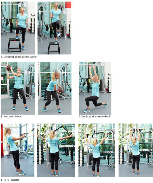
McGill’s first two steps for training the abdominals involve teaching clients how to maintain stability of the spine and training efficient movement patterns. Once clients can consciously maintain stability of the spine, it is important to have them work from a standing position. One strategy, for example, is to include single-leg balance exercises, which can help integrate abdominal function with the lower extremities. Other strategies include recreating the movements of gait and having the rib cage and pelvis rotate over one another, or having the spine remain in a lengthened position as one leg moves into extension. Yet another strategy is to have the client maintain stability of the spine while moving the arms and legs. There are a variety of ways to engage the muscles that control the center of gravity (Table 2)—just make sure you first understand the skills and abilities of your clients or class participants before you determine which strategies to use in your program design.
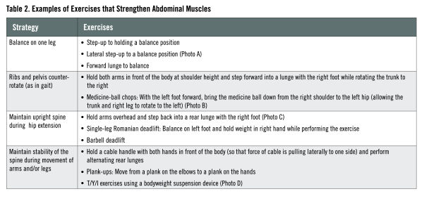
The abdominal muscles contain a combination of type I and type II fibers. Therefore, low-intensity, long duration stabilization exercises can be used one day to activate the type I fibers, while heavy resistance strength or power exercises can be used the next day to stimulate the type II fibers. Strength-training exercises such as the barbell deadlift, Romanian deadlift and Turkish get-up can be extremely effective for enhancing the strength of all the layers of the abdominal muscles.
Despite how many integrated core exercises you include in a training session, many clients and class participants will still expect to perform traditional crunches so they can check “core training” off their exercise to-do lists. To avoid this awkward interaction, you can always include a few crunches at the end of a workout or class to give them what they want, which, ultimately, will help keep them coming back.
References and Recommended Reading
Abernathy, B. et al. (2005). The Biophysical Foundations of Human Movement (2nd ed.). Champaign, Ill.: Human Kinetics.
Bergmark, A. (1989). Stability of the lumbar spine: A study in mechanical engineering. Acta Orthopaedica Scandinavica Supplementum, 230, 60, 20-29.
Contreras, B. and Schoenfeld, B. (2011). To crunch or not to crunch: An evidence-based examination of spinal flexion exercises, their potential risks and their applicability to program design. Strength and Conditioning Journal, 33, 4, 8-18.
Enoka, R. (2002) Neuromechanics of Human Movement (3rd ed.). Champaign, Ill.: Human Kinetics.
Hibbs, A. et al. (2008). Optimizing performance by improving core stability and core strength. Sports Medicine, 38, 12, 996-1008.
McGill, S. (2010). Core training: Evidence translating to better performance and injury prevention. Strength and Conditioning Journal, 32, 3, 33-46.
McGill, S. et al. (2009). Exercises for the torso performed in a standing posture: Spine and hip motion and motor patterns and spine load. The Journal of Strength and Conditioning Research, 23, 2, 455-464.
McGill, S., Karpowicz, A. and Fenwick, C. (2009). Ballistic abdominal exercises: Muscle activation patterns during three activities along the stability/mobility continuum. The Journal of Strength and Conditioning Research, 23, 3, 898-905.
McGill, S., Karpowicz, A. and Fenwick, C. (2009). Comparison of different strongman events: trunk muscle activation and lumbar spine motion, load and stiffness. The Journal of Strength and Conditioning Research, 23, 4, 1148-1161
Myers, T. (2014). Anatomy Trains (3rd ed.). London: Elsevier.
Neumann, D. (2010). Kinesiology of the Musculoskeletal System: Foundations for Rehabilitation (2nd ed.). St. Louis, Mo.: Elsevier.
Schleip, R. et al. (2012) Fascia: The Tensional Network of the Human Body. London: Churchill Livingstone.
 by
by